What Is Age-related Macular Degeneration

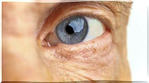
Age-related macular degeneration, also called age spots, is an eye disorder characterized by gradual acute and central vision loss. This makes it increasingly difficult to visualize fine details and to read.
This disorder is called age-related macular degeneration (AMD) because this pathology is significantly more common in people over 50 years of age. Usually the process happens slowly over time, but sometimes it progresses faster.
Currently, several studies are underway to correct macular degeneration in relation to age. In addition, progress has been made in the genetic understanding of the disease. There are also promising studies related to healthy cell transplantation to correct the problem.
What is age-related macular degeneration?

Specifically, age-related macular degeneration has to do with a problem with the retina. This tissue is sensitive to light and it is located at the base of the eye. Its function is to convert light and images into electrical impulses that are transmitted to the brain.
The macula is located in the center of the retina. It is a yellow spot whose function is to capture the finest details in sight. When this disorder occurs, the central vision worsens, but peripheral vision remains the same.
This disease is more common in people with tobacco addiction and people who are overweight. In addition, it is more common in those who suffer from high blood pressure, high cholesterol levels and diabetes.
Also read: 7 early symptoms of diabetes
Types of age-related macular degeneration
There are two types of age-related macular degeneration: wet and dry. Wet macular degeneration occurs when blood vessels grow abnormally under the retina. Such vessels are fragile and tend to drip blood and fluid. All this causes the macula to rise from its normal position. This damages the macula and it will break down quickly.
The wet type is the most severe of the macular degenerations. One of the first symptoms is to see straight lines as if they were wavy.
Dry macular degeneration, on the other hand, is the most common form of this pathology. It occurs when macula cells that are sensitive to light begin to deteriorate. Vision becomes blurred and it becomes increasingly difficult to maintain central vision.
It develops in three phases:
- Early macular degeneration. To begin with, the person does not experience any symptoms. However, when examining the retina, the doctor will notice yellow deposits called the drusen under the retina.
- Moderate AMD. Then the patient will have many drusers or large drusers. They may have blurred vision or they may need more light to read.
- Advanced macular degeneration. Finally, in this stage there are many drusen and there will also be cell and tissue deterioration in the central area of the retina. The patient’s central vision is opaque and blurred. In addition, they will have difficulty reading and recognizing faces.
Causes
Experts are still not sure why age-related macular degeneration occurs. It obviously has to do with the natural deterioration of the body, related to the passage of time. In addition, however, there is not much information.
What we do know is that there are risk factors that can predict this disease. The most important are:
- Family history
- Smoking
- Nutritional factors such as diet rich in fat
- To be white
- To be a woman
Also read: 5 Natural Remedies That Can Stop Macular Degradation
Diagnosis and prognosis

Generally, age-related macular degeneration is detected through an eye examination. The test typically includes the following test:
- Visual acuity. This measures the patient’s vision at different distances.
- Tonometry measures the pressure inside the eyes using a special device.
- Pupil dilation. The physician pours drops into the patient’s eyes to dilate the pupil. The doctor will then examine the retina and optic nerve with a magnifying lens.
- Amsler card. This card has a grid that looks like a chessboard. The patient covers one eye and looks at a black spot in the middle of the grid. Then repeat the exercise with the other eye.
The prognosis for dry macular degeneration is generally positive. Usually, vision loss is not devastating. On the other hand, in wet macular degeneration, there is often a strong loss of vision. In this case, the patient will not be able to perform activities that require visualization of fine details.
Lastly, if you notice any of the symptoms we have mentioned, do not hesitate to seek out a specialist who will help treat it in the best possible way.